Human pluripotent stem cells (hPSC): 2D culture versus in vivo
Standard 2D hPSC culture consists in seeding hPSCs as single cells or, most frequently, cell clusters in culture flasks or Petri dishes coated with extracellular matrix. Cells are then incubated at 37°C under normoxic or hypoxic conditions. Every 24-48 hours, hPSCs are taken out of the incubator for media change. When the colonies approach or reach confluence, the cell monolayer is detached from the substrate, often resulting in a combination of cell clusters and single cells, which are then replated for further expansion.
The first obvious limitation of 2D culture for the recapitulation of the in vivo environment of hPSCs is the intermittent control of cell culture parameters, including temperature, pH, dissolved oxygen (DO), glucose, and growth factor concentrations, caused by medium changes.
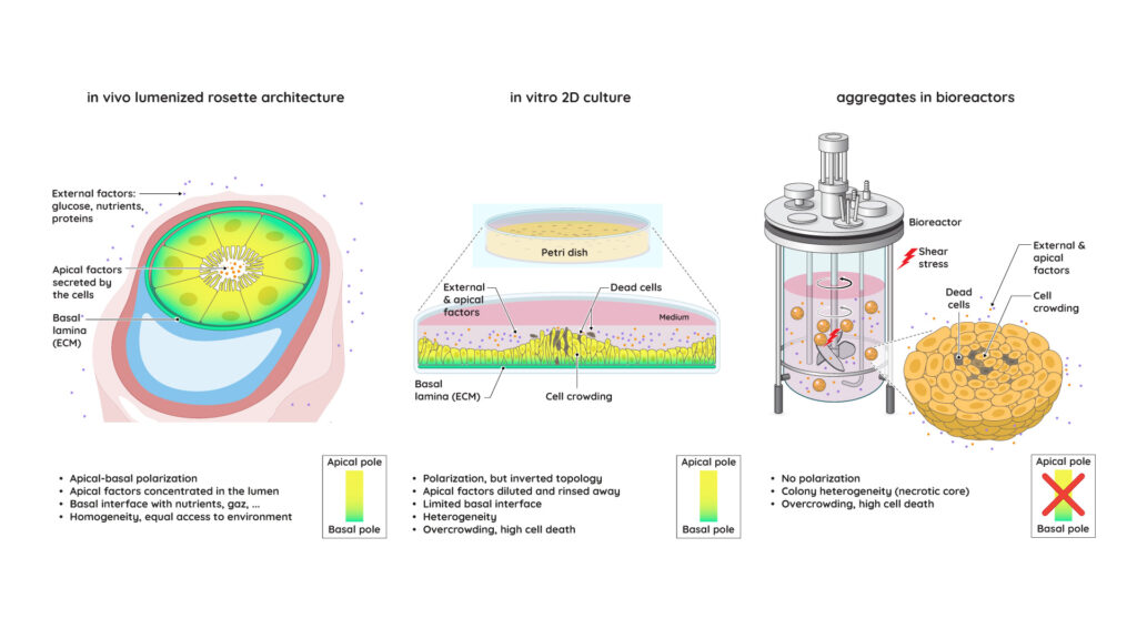
Second, while it has been reported that hPSCs cultured in 2D spontaneously form lumens, such rosette-like structures were found to rapidly collapse (in ~5 days or less). hPSCs end up establishing an inverted topology in 2D. As opposed to the rosette architecture, in Petri dishes the apical domain is facing the cell culture media, while the basal pole is established at the bottom of the dish, with limited access to external cues and nutrients. As a result, in 2D culture, signalling molecules secreted at the apical domain are diluted in a large volume of media, which is rinsed at every media change. Developmental biologist Marta Shahbazi estimated a 2,500-fold difference in the concentration of autocrine factors in the lumen of hPSC colonies in rosette conformation versus cell culture medium in standard 2D culture.
Furthermore, in contrast with the lumenized rosette conformation, in 2D, as the colony expands and reaches confluence, cells at the center of the colony experience a very different environment from the ones at the edge, translating into spatially heterogeneous expression of pluripotency markers. This phenomenon is described as edge/center heterogeneity. The cellular crowding and compaction observed in 2D hPSC colonies also induce a competition for space, known to inhibit epithelial cell proliferation and trigger apoptosis through caspase-dependent mechanisms. This lateral competition is independent of cellular fitness markers and promotes the selection of fast-growing apoptotic-resistant clones, likely to harbor oncogenic mutations.
Together, intermittent control of cell culture conditions, unphysiological apico-basal polarity, edge-center heterogeneity, and competition for space may explain the high mortality rates, spontaneous differentiation, and genetic drift reported in 2D hPSC colonies, highlighting the limitations of monolayer cultures for scaling up the production of clinical-grade hPSC-derived products.

Fig. Comparison of standard 2D culture and 3D aggregate culture to the in vivo lumenized rosette
Pluripotent stem cells: 3D aggregates in bioreactors versus in vivo
With the aim of meeting industrial needs for scale-up, hPSCs are also cultured in agitated bioreactors, which provide continuous control over key cell culture parameters. In standard bioreactor culture, agitation induces the formation of 3D aggregates of hPSCs. However, productivity and cell quality are consistently found to be lower in this configuration than in 2D. In a benchmark presented in 2021 at the American Society of Gene & Cell Therapy (ASGCT), prolonged agitated hiPSC culture over 28 days resulted in a 6,000-fold lower expansion than in 2D, and a higher mutational load was reported in hPSC aggregates.
Several factors may affect performance in bioreactors. In vivo, an extra-embryonic tissue layer protects hPSCs from external stress. In bioreactors, hPSCs aggregates are directly exposed to impeller-induced hydrodynamic stress, which can trigger either cell death or spontaneous differentiation. Agitation also leads to spontaneous fusions of aggregates, resulting in large hPSC spheroids harboring necrotic cores instead of lumens. Cellular overcrowding, heterogeneous access to gas, nutrients, and signalling molecules in large hPSC aggregates, together with failure to establish apical-basal polarity, and epiblast-like cell competition, may altogether explain the reduced proliferation and increased rate of mitotic errors observed in bioreactor culture.


