

Human primed pluripotency: a 7-day window
Pluripotency is a transient cell property in vivo. At the end of the first week of development, naïve hPSCs transition to primed pluripotency. Within the following 5 to 7 days, the hPSC colony, benefiting from a very fast 15h to 16h cell cycle, grows exponentially to reach over 1,000 cells. Following this exponential amplification, hPSCs exit pluripotency as they commit to one of the three germ layers that undergo gastrulation. Overall, among the 40 cell divisions required to constitute an adult body (~37 trillion cells), 10 are estimated to occur within the second week of development, highlighting the importance of the pluripotent stage.
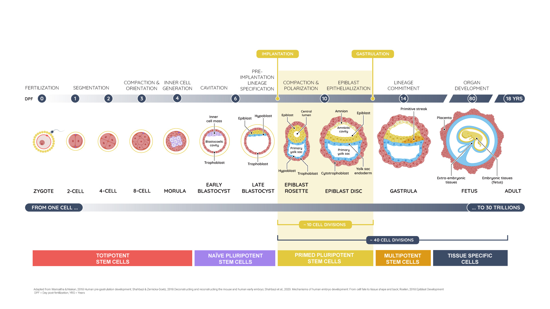
Human primed pluripotency in vivo: a 7 day-window, a protected microenvironment, and a 3D epithelial organization.
A protected microenvironment
Before implantation, the fertilized egg and its subsequent totipotent stem cell progeny grow for a few days within the zona pellucida – a 5-10 μm thick spherical shell composed of glycoproteins. The zona pellucida is a mechanically protective capsule that allows for metabolic exchanges with the environment of the uterus. This structure is hatched around day 7 post-fertilization to enable implantation into the uterine wall. This capsule is then functionally replaced by an extra-embryonic outer layer of trophoblast cells, which provides mechanical protection, enables metabolic exchanges, and creates a controlled 3D niche in which hPSCs can grow exponentially.
A distinctive rosette architecture
After implantation, hPSCs acquire primed pluripotency and arrange as a spherical single cell-layered epithelium organized around a central cavity. This lumenized rosette architecture is instrumental for the in vivo function of the hPSC compartment.
First, this radial organization around a central lumen ensures that each hPSC within the colony has similar environmental conditions, including topology, mechanical characteristics and access to oxygen, nutrients, extracellular matrix, and growth factors. Its radial symmetry per se avoids local variations, which could compromise the establishment of ontogenic axes organizing the body plan.
Second, in the epiblast rosette, all hPSCs have their apical domains clustered toward the lumen, which acts as central signalling hub, enabling synchronous and homogeneous growth.
Finally, the lumenized rosette is a plastic architecture suited for growth. Its roundness suggests a positive intraluminal osmotic pressure. Such a pressure gradient may help expand the surface area of the epithelium as hPSCs divide, and locally decrease the tangential mechanical resistance to accommodate new cells. Facilitating cell division, this conformation also prompts the depletion of abnormal cells, thus safeguarding the genomic integrity of the hPSC colony.

